
Microtia
Loading

Loading

A wide range of patients with microtia (15–60%) present with additional abnormalities. The most frequent simultaneous dysmorphic features associated with microtia are facial asymmetry, cleft palate, macrostomia and epibulbar dermoids. In addition, complete or partial facial paralysis can be observed in 20% of the patients.
| LOCAL PROBLEMS |
|---|
| Low Hairline, Ectopic Ear Remnants, Skin Tags, Dermoid Sinus, Cysts |
| ANOMALIES RELATED WITH THE OPPOSITE EAR |
|---|
| Prominent Ear Deformity, Cup Ear, Ear Lobe Deformities, Skin Tags |
| OROFACIAL ANOMALIES |
|---|
| Hemifacial Microsomia (HFM), Congenital Facial Palcy, Mandibular Hipoplasia, Macrostomia, Cleft Palate etc. |
| EYE PROBLEMS |
|---|
| Coloboma, Microphtalmia, Epibulbar Tumors etc. |
| SYSTEMATIC PROBLEMS |
|---|
| Vertebrocostal Anomalies, Renal Anomalies, Cardiac Anomalies |
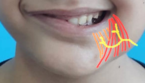
Complete or partial facial paralysis is a condition that often accompanies microtia. Asymmetry in facial expressions is manifested by signs such as a shift of the mouth when crying, laughing, or the inability of the eyelids to close completely. We can also perform some interventions to correct facial paralysis during ear construction within the same surgery.
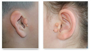
These embryonal remnants, which are located in the microtic ear or the opposite ear and contain skin, fat and sometimes cartilage tissue, do not pose any health problems, but they can be removed during ear surgery to visually normalize the patient.
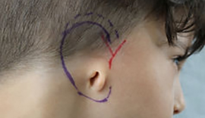
A low hairline is seen in approximately 10% of microtia cases. This is an important factor affecting our decision in choosing the methods and approaches to be used in ear reconstruction. If there is hair on where we will positionate the new ear, our preference is to remove hair from the area with laser epilation before ear reconstruction.
In cases of unilateral microtia, it is common to see deformities such as prominent ear, cup ear, etc. in the opposite ear. Depending on the type and severity of the deformity, it is possible to correct these deformities observed in the opposite ear either during the ear reconstruction stages or after the auriclar reconstruction is completed.

Only 10% of microtia patients are syndromic. These are cases where microtia is seen with additional anomalies. The most common congenital anomalies accompanying microtia are Hemifacial Microsomia, Goldenhar and Treacher Collins syndromes.
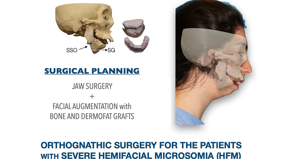
As a result of the influence on the facial elements that develop from the same embriological origin as the ear during the development process in the womb, facial asymmetry is observed in the cheekbones, cheeks and chin due to developmental deficiency. Their treatment can be carried out with dermofat graft and/or maxillofacial surgery, which can be performed simultaneously with auricular reconstruction depending on the severity of the anomaly.

The most prominent manifestation of Goldenhar Syndrome is microtia and hemifacial microsomia. In addition to these findings, there are benign tumors of the eye (epibulbar dermoids). Children with Goldenhar syndrome may also have neck problems caused by adhesions or bone bridges between the neck vertebrae.
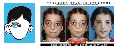
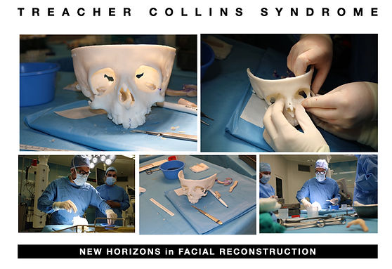
Treacher Collins syndrome (TCS), which people all over the world become aware of with the movie entitled "WONDER", is a genetic disorder characterized by deformities of the ears, eyes, cheekbones, and chin. The degree to which a person is affected, however, may vary from mild to severe. Complications may include breathing problems, problems seeing, cleft palate, and hearing loss. It is a complex anomaly whose treatment requires special expertise and experience.

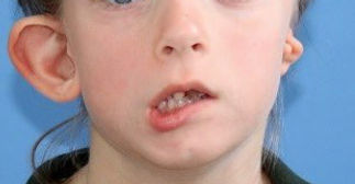
Have you noticed an involuntary shift in your childs lower lip when crying or laughing? This is a common problem in children with microtia and occurs due to the marginal mandibular nerve palsy, a branch of the facial nerve. We can correct this problem with a surgical procedure that can be applied in the first stage of ear reconstruction.
| CORRECTION OF ASYMMETRICAL SMILE |
|---|
|
Have you noticed an involuntary shift in your childs lower lip when crying or laughing? This is a common problem in children with microtia and occurs due to the marginal mandibular nerve palsy, a branch of the facial nerve. We can correct this problem with a surgical procedure that can be applied in the first stage of ear reconstruction.
 |
| FAT GRAFTING FOR HEMIFACIAL MICROSOMIA |
|---|
|
In the second stage, which is carried out at least 6 months after the first surgery, the constructed auricle is lifted to create the posterior sulcus of the auricle. During this surgery, the facial deformity of the patients who have facial asymmetry due to Hemifacial Microsomia (HFM) can be corrected with filling the face with patients own tissue. The fat, skin and spare cartilage necessary for this procedure are removed from the same incision to avoid leaving additional scars. There will be no drains and pain in the chest area. The patient can usually be discharged from the hospital 2 days after surgery.
 |
